Real-time Brain Scans During Ketamine Therapy: Insight & Evidence

August 7, 2025
Understanding Neurophysiological and Neuroimaging Insights into Ketamine Therapy
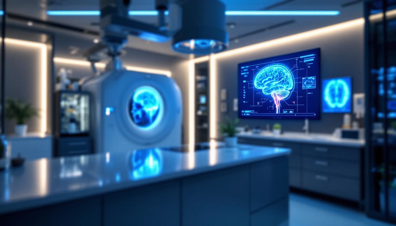
Recent advancements in neuroimaging techniques have shed light on the complex neural mechanisms underlying ketamine therapy. By capturing real-time brain activity, scientists are uncovering how this innovative treatment influences neural circuits, promotes neuroplasticity, and induces altered states of consciousness. This comprehensive analysis explores these findings in depth, emphasizing the role of structural and functional changes in the brain during and after ketamine administration.
Neurophysiological Effects of Ketamine on the Brain During Therapy
What are the neurophysiological effects of ketamine on the brain during therapy?
Ketamine has profound effects on brain activity that help explain its rapid antidepressant and dissociative properties. During treatment, it modulates the balance of neural excitation and inhibition, particularly within the prefrontal cortex, which is involved in mood regulation and cognition. Studies utilizing neuroimaging techniques like EEG and fMRI reveal that ketamine increases gamma oscillatory power, which signifies more synchronized neural firing. Such enhanced synchronization supports the rapid reorganization of brain circuits necessary for its therapeutic effects.
Additionally, ketamine influences brain signal entropy, reflecting a more complex and diverse pattern of neural activity. This increased entropy correlates with altered consciousness and psychedelic states experienced during therapy. EEG recordings also show a decrease in low-frequency power, such as delta waves, suggesting a shift towards higher-frequency oscillations. These shifts are associated with heightened cortical excitability and might facilitate synaptic plasticity.
Neural oscillations, particularly gamma waves, are known to play a critical role in cognitive processes and emotional regulation. During ketamine infusion, gamma activity increases, supporting the idea that ketamine enhances neural plasticity and connectivity. At higher doses, effects extend to slow delta waves, which are typically linked to deep sleep but are observed here to be involved in the drug’s dissociative and sedative effects.
Furthermore, ketamine impacts regions involved in emotional processing, often reducing activity in the amygdala and anterior insula. These areas are crucial for attaching emotional significance to stimuli and are hyperactive in depression. By dampening their activity temporarily, ketamine helps alleviate depressive symptoms and dissociative experiences.
Overall, the neurophysiological changes—ranging from oscillatory shifts to network reconfiguration—are central to ketamine's ability to induce rapid antidepressant responses and transient altered states. This dynamic modulation of brain activity underscores its potential as a powerful tool in mental health treatment, offering insights into both its therapeutic benefits and mechanisms of action.
Neuroimaging Evidence of Brain Activity Modulation

How does ketamine influence brain activity as observed through neuroimaging studies?
Neuroimaging research provides valuable insights into how ketamine affects the brain's structure and function, especially in the context of depression and altered states of consciousness. Using advanced techniques like functional magnetic resonance imaging (fMRI) and diffusion MRI, scientists have observed multiple changes in brain connectivity and activity following ketamine administration.
One prominent effect is the modulation of large-scale brain networks. Ketamine reduces activity in limbic and emotion-related regions such as the amygdala and subgenual anterior cingulate cortex (sgACC). These areas are crucial in emotional processing and mood regulation, and their hyperactivity is often linked to depressive symptoms. A decrease in blood flow and activity in these regions correlates with clinical improvements, highlighting ketamine’s rapid antidepressant effects.
Conversely, ketamine enhances activity in regions involved in mood regulation and cognitive functions, including the prefrontal cortex, hippocampus, and thalamus. These areas are associated with executive function, memory, and sensory processing. Increased connectivity between these regions has been observed, supporting the drug’s role in promoting neural plasticity.
Furthermore, ketamine disrupts connectivity within large-scale networks like the default mode network (DMN) and salience network (SN). Often, these networks show decreased functional connectivity, which is thought to reduce maladaptive rumination and emotional dysregulation common in depression.
Structural brain changes have also been documented. White matter microstructure alterations, such as increased diffusivity in certain tracts, suggest that ketamine promotes neuroplasticity. Notably, hippocampal volume can transiently increase, reflecting ongoing neurogenesis or synaptic growth, which supports mood improvement.
Finally, the brain-wide impact of ketamine extends to neurochemical systems, including dopamine networks. Repeated ketamine use can lead to long-term structural modifications, affecting dopamine pathways related to motivation and reward.
In summary, neuroimaging evidence demonstrates that ketamine influences brain activity by decreasing hyperactive limbic circuits and enhancing the function of prefrontal, hippocampal, and thalamic regions. These changes facilitate improvements in mood and cognition, underpinning the drug's rapid antidepressant and psychotropic effects.
Neural Mechanisms Influenced by Ketamine Therapy

What do neuroimaging studies reveal about the neural mechanisms affected by ketamine therapy?
Recent brain imaging research provides valuable insights into how ketamine exerts its rapid antidepressant effects. Using advanced techniques like functional magnetic resonance imaging (fMRI) and high-resolution MRI scans, scientists have observed how ketamine influences brain activity and connectivity.
Ketamine appears to modulate key brain circuits involved in mood regulation and emotional processing. It reduces activity and connectivity in the default mode network (DMN) and self-monitoring regions such as the medial prefrontal cortex. Concurrently, it increases activity in areas linked to reward and pleasure, including the striatum, which may help alleviate depressive symptoms quickly.
One notable finding is ketamine's impact on the amygdala, hippocampus, and the anterior insula—regions critical in emotion regulation. After treatment, there is often a decrease in activity within the subgenual anterior cingulate cortex (sgACC), a region heavily involved in depression. Changes in connectivity between the prefrontal cortex and limbic structures suggest improved emotional regulation.
Beyond functional activity, ketamine affects physical brain properties. Studies have shown altered cerebral blood flow, increased glucose metabolism, and enhancements in white matter integrity, indicating broad neuroplastic effects.
Specifically, the modulation of neural circuits involves decreased connectivity within the DMN, which aligns with reduced rumination and self-referential thought. Conversely, increased activity in reward-related circuits may underpin the rapid mood improvements observed post-infusion.
Overall, neuroimaging studies demonstrate that ketamine's therapeutic action involves a complex rebalancing of brain networks responsible for mood, emotion, and cognition. These modifications promote faster symptom relief and pave the way for targeted treatments that harness brain plasticity.
Altered States and Consciousness in Ketamine Therapy

How is brain activity during ketamine treatment related to changes in consciousness and altered states?
During ketamine therapy, distinct patterns of brain activity correspond with various altered states of consciousness. Neuroimaging studies reveal that ketamine influences neural circuits involved in affective processing, self-awareness, and multisensory integration.
Ketamine induces dose-dependent changes in brain oscillations, particularly in the alpha and gamma frequency bands. Typically, there is a reduction in alpha power, especially in regions like the precuneus and temporoparietal junction (TPJ), which are critical for self-referential thought and perceptual integration. Conversely, gamma activity often increases, correlating with heightened perceptual vividness and dissociative experiences.
These electrophysiological changes are closely tied to subjective states such as depersonalization, ego transcendence, and derealization. For example, decreased alpha oscillations in the TPJ are linked to feelings of disembodiment, while increased gamma oscillations may underlie the complex visual and perceptual phenomena reported during treatment.
Ketamine’s mechanism involves blocking NMDA receptors, which leads to disinhibition of excitatory neurons. This disruption of cortical communication, particularly in networks integral to multisensory integration and self-perception, results in altered states.
Functional MRI and EEG studies show that during ketamine administration, there is decreased activity in regions responsible for maintaining a stable sense of self, such as the posterior cingulate cortex, and increased activity in areas integrating sensory information. These neural dynamics contribute to the dissociative states characteristic of ketamine's psychedelic effects.
Moreover, the degree of neural oscillation disruption and the intensity of altered states may be predictive of treatment success. Some research suggests that more profound dissociative experiences, linked with greater EEG changes, correlate with better antidepressant responses.
In summary, brain activity during ketamine treatment reflects a complex interplay of oscillatory modifications and network disconnections that facilitate extraordinary conscious experiences. Understanding these neurophysiological patterns deepens our knowledge of how altered states can mediate therapeutic effects and informs future clinical applications.
Neuroplasticity and Long-term Brain Changes
What role does neuroplasticity play in the therapeutic effects of ketamine?
Neuroplasticity is fundamental to how ketamine exerts its rapid antidepressant effects. By promoting the growth and remodeling of synapses, ketamine enhances the brain's capacity to reorganize and adapt, particularly in regions crucial for mood regulation such as the prefrontal cortex, amygdala, and hippocampus. These structural changes include increases in dendritic spine density and synaptic connectivity, which help restore normal brain function.
One of the key processes involved is the release of brain-derived neurotrophic factor (BDNF), a protein that supports neuron survival and synaptic growth. Ketamine activates TrkB receptors, which are critical for synaptic formation and strengthening. This activation encourages the formation of new synapses, thereby contributing to fast and sustained improvements in depressive symptoms.
Research shows that these neuroplastic changes are dose-dependent and mediated by specific neurotransmitter systems. For instance, activation of dopamine D1 receptors and enhanced AMPA receptor activity are involved in the process. The effects on neuroplasticity are typically observed within hours of administration, creating a “critical window” where the brain is more receptive to psychotherapy and behavioral interventions, potentially leading to more lasting therapeutic benefits.
In summary, ketamine’s ability to stimulate synaptic growth and foster neural connectivity underpins its rapid therapeutic action, setting the stage for long-term recovery from depression by fundamentally reshaping brain circuits.
| Aspect | Description | Additional Notes |
|---|---|---|
| Synaptic growth | Increase in dendritic spines and synaptic connections | Supports neural communication and learning |
| BDNF release | Elevated following ketamine, promotes synaptic plasticity | Critical for neuron health and growth |
| TrkB activation | Receptor activation essential for neuroplastic effects | Facilitates formation of new synapses |
| Dose dependence | Effects vary with dosage, influencing the extent of plasticity | Informs optimal treatment protocols |
| Critical intervention window | Period of heightened plasticity post-treatment | Enhances potential for psychotherapy |
This neuroplasticity not only explains the rapid mood improvements seen with ketamine but also offers a mechanistic basis for combining ketamine treatment with therapies like psychotherapy, maximizing therapeutic outcomes by leveraging this brief window of heightened brain flexibility.
Implications for Therapy and Future Research

How can ketamine therapy be combined with psychotherapy during the neuroplasticity window?
The neuroimaging studies reveal that ketamine induces a surge in brain plasticity, particularly within the first 24 to 48 hours after administration. This period represents a critical window where the brain is more receptive to change. Clinicians can capitalize on this by scheduling psychotherapy sessions during this optimal timeframe. Engaging patients in therapy while their brain networks are highly adaptable could potentially lead to faster, more durable mental health improvements, especially for treatment-resistant depression.
How are advanced imaging techniques paving the way for personalized treatment approaches?
High-resolution MRI scans, including 3T and 7T MRI, alongside functional neuroimaging like fMRI, are revolutionizing our understanding of individual neurobiological responses to ketamine. These tools allow scientists to observe real-time changes in brain structure and activity, identifying specific circuits involved in each patient’s condition. Such detailed insights open the door for tailoring treatments based on a patient's unique brain profile, enhancing outcomes and reducing side effects.
What developments are underway in creating targeted interventions using neuroimaging biomarkers?
Progress in identifying biomarkers such as reductions in SV2A for Alzheimer’s or dopamine network alterations in ketamine users guides the development of precise interventions. By using neuroimaging data, researchers aim to craft targeted treatments that modify specific brain pathways implicated in disorders. This approach could lead to the creation of new drugs or stimulation techniques optimized for individual neural architectures, ultimately advancing personalized medicine in neuropsychiatry.
| Aspect | Current Advances | Future Directions |
|---|---|---|
| Imaging Techniques | 3T/7T MRI, fMRI, PET | Multi-modal imaging combining structural and functional data |
| Treatment Personalization | Brain circuit profiling | Real-time monitoring and adaptive treatment strategies |
| Biomarker Development | SV2A, dopamine receptor changes | Reliable biomarkers for early diagnosis and treatment response |
Focusing on these areas will improve understanding and treatment of complex brain disorders, making neuroimaging an integral part of future mental health care.
Unlocking the Brain's Response to Ketamine
As neuroimaging technologies continue to evolve, our understanding of how ketamine modulates brain activity, connectivity, and plasticity deepens. These insights not only elucidate the neurobiological basis of its rapid antidepressant effects but also pave the way for more personalized, targeted therapeutic strategies. By harnessing real-time brain scans, clinicians can optimize treatment protocols, monitor neural responses, and advance the development of novel interventions for depression, PTSD, and other neuropsychiatric disorders.
References
- Unveiling the Brain's Response to Ketamine in Depression
- Ketamine's acute effects on negative brain states are mediated ...
- Subanesthetic Ketamine Treatment Promotes Abnormal Interactions ...
- First evidence for higher state of consciousness found | Imperial News
- Brain Changes Associated With Long-Term Ketamine Abuse, A ...
- Imaging Analysis Suggests How Ketamine Treatment Might Have ...
- Transcranial magnetic stimulation and ketamine - Frontiers
Recent Posts
Conditions Treated
AnxietyDepressionOCDPTSDPostpartum DepressionPain ManagementSubstance AbuseSuicidal IdeationOur Location